- Research Services
- Capabilities
- About Us
- Resources
- Contact Us
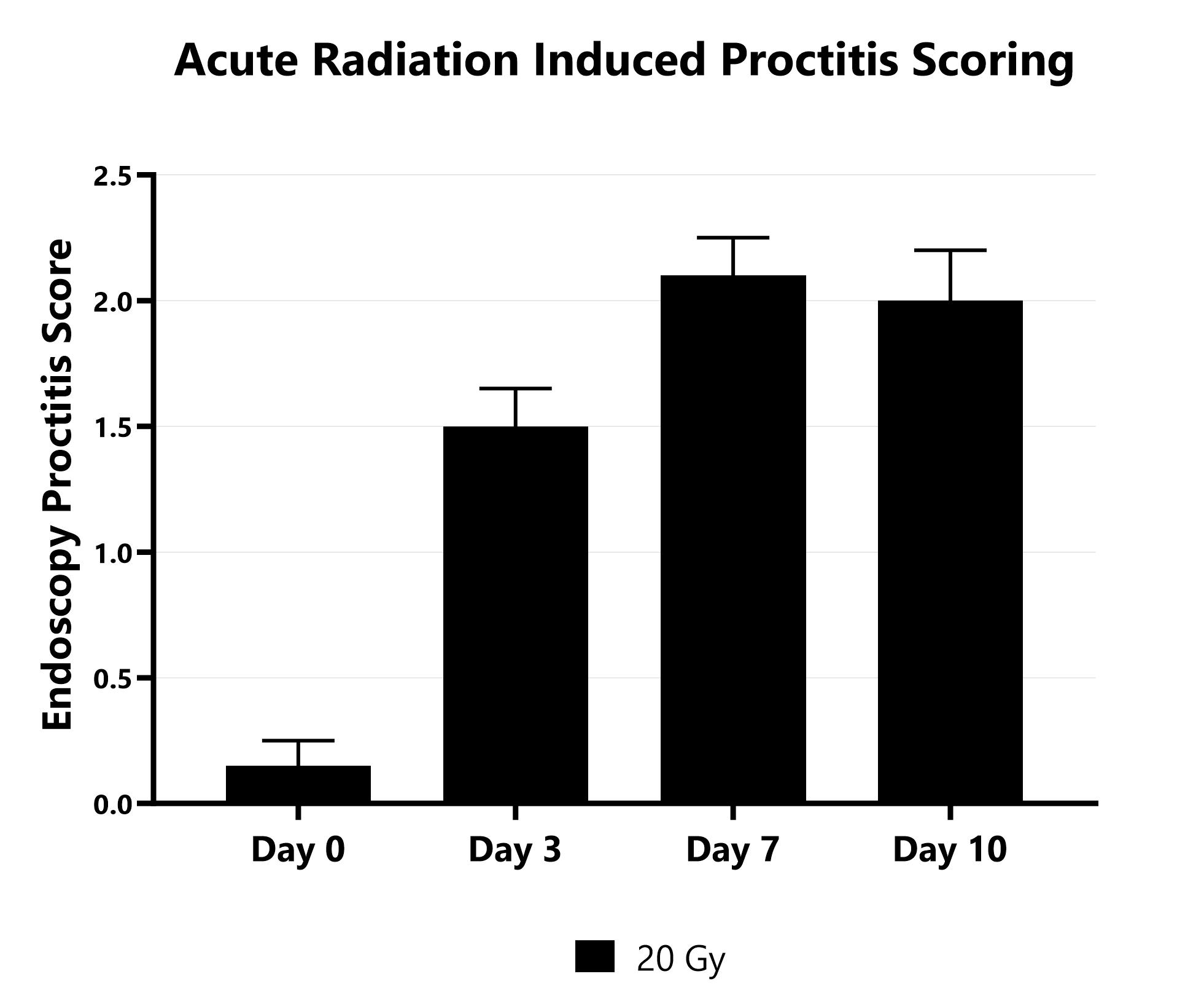
Proctitis severity is assessed longitudinally using video endoscopy at multiple timepoints during an acute radiation-induced proctitis study.
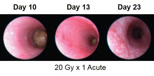
Proctitis severity is assessed longitudinally using video endoscopy at multiple timepoints during an acute radiation-induced proctitis study.
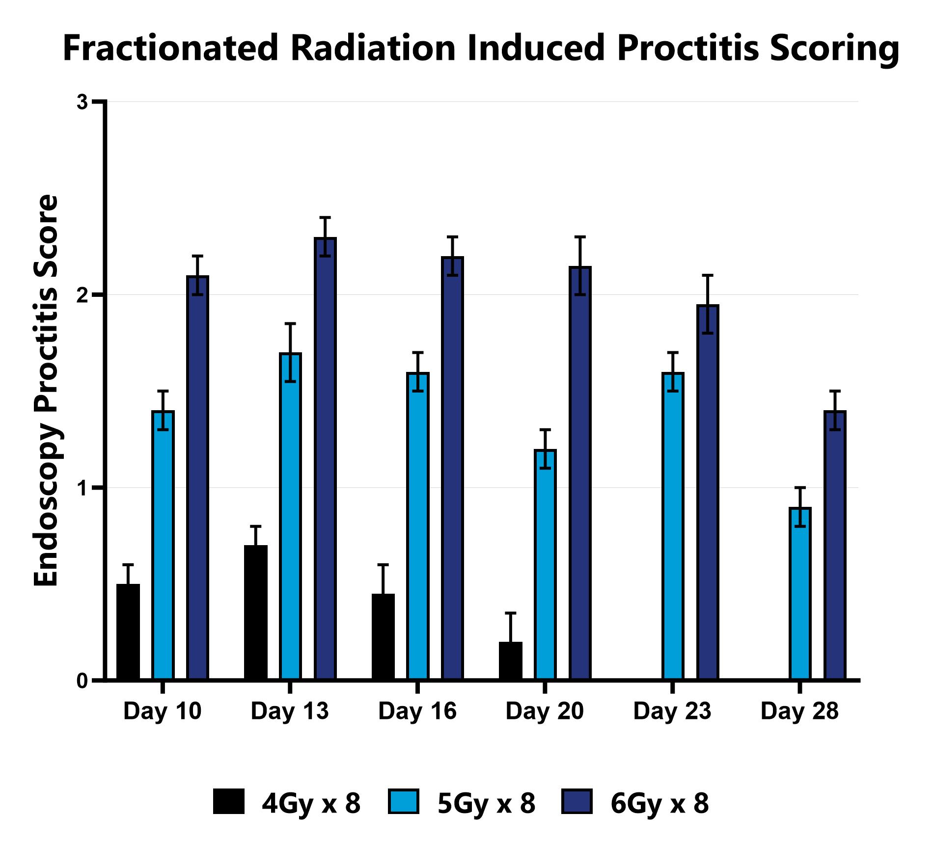
Proctitis severity is assessed longitudinally using video endoscopy at multiple timepoints during a fractionated radiation-induced proctitis study.
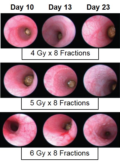
Proctitis severity is assessed longitudinally using video endoscopy at multiple timepoints during a fractionated radiation-induced proctitis study.
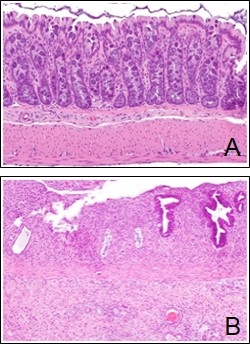
Following fractionated radiation treatment, rectal tissue is collected. (A) Photomicrograph showing histological appearance of normal rectal mucosa. (B) Photomicrograph showing histological appearance of inflamed rectal mucosa.